Technology

Rethinking how light reaches the retina.

Our technology is built around our patented trans-pars-planar illumination — an approach to enable ultra-widefield, non-mydriatic imaging of the retina without splitting the pupil.

The approach

Trans-pars-planar illumination.

Constructing a wide field fundus camera is technically challenging because conventional systems send illumination and imaging through the same narrow pupil path. We take an alternative approach in which light is delivered through the pars plana, separating illumination from the imaging path, up to 200 degrees internal eye angle.

Ultra-widefield imaging

By separating illumination from imaging, the approach is being developed to capture a wide field of the retina.

Non-mydriatic

A central design goal of the program is examination without the need for pharmacologic pupil dilation.

Portable by design

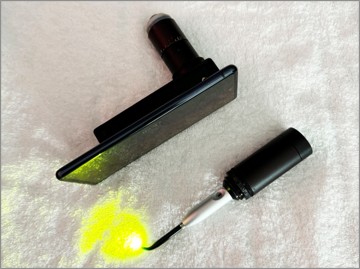
The systems are being developed to be portable and easy to use, including a smartphone-based configuration.

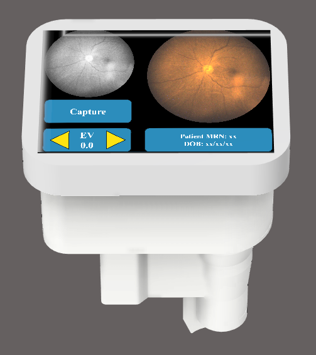
PedCam device with imaging interface
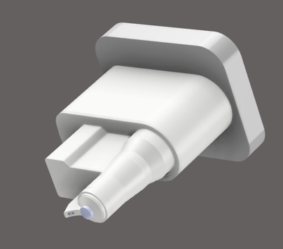
PedCam device render
Products under development

PedCam

A dedicated handheld ultra-widefield fundus camera for non-mydriatic examination of the pediatric retina, including applications in retinopathy of prematurity.

  • Ultra-widefield

    Trans-pars-planar illumination for border-to-border viewing, beyond 200 degrees.

  • Non-mydriatic

    Designed for examination without pharmacologic pupil dilation.

  • Portable, handheld form

    Intended for use at the point of care.
Products under development

PedCam-SP

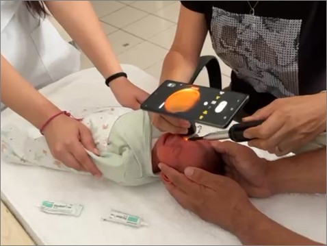
A smartphone-based ultra-widefield imaging configuration, intended to make comprehensive pediatric retinal examination more portable and accessible.

  • Ultra-widefield

    Trans-pars-planar illumination principle for border-to-border viewing.

  • Non-mydriatic

    Designed for examination without pharmacologic pupil dilation.

  • Smartphone-based

    Built around widely available mobile hardware.

PedCam-SP in use
PedCam-SP hardware
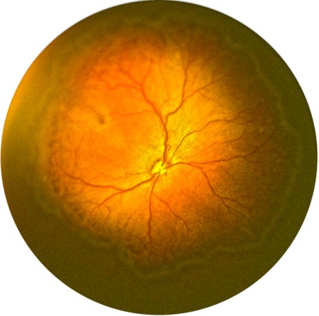
Example images

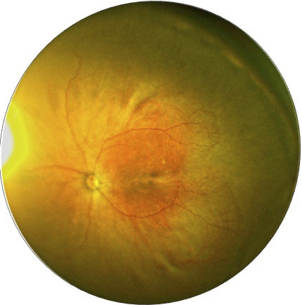
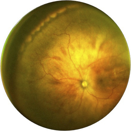
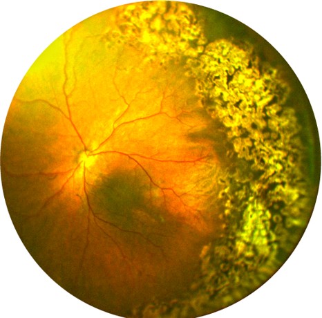
Representative ultra-widefield retinal images captured by existing prototypes

See the research behind the program.

Review our NIH SBIR record and publications.

Research & funding